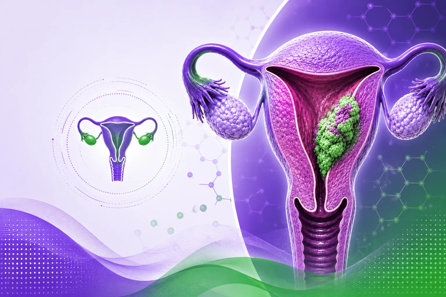
Corpus uteri cancer, also known as uterine cancer, occurs when abnormal cells develop in the uterus. It includes endometrial cancer, which begins in the inner lining of the uterus, and uterine sarcoma, which develops in the muscle wall. Precision oncology and genomic profiling help guide personalized treatment decisions for improved patient care.
Corpus uteri cancer is the abnormal growth of cells in the uterus. It includes endometrial cancer, which develops in the lining of the uterus, and uterine sarcoma, which develops in the muscle wall of the uterus.
Early diagnosis combined with precision oncology helps identify molecular changes that guide personalized treatment strategies and improve outcomes.
Early diagnosis and comprehensive molecular testing help determine the stage of disease and guide individualized treatment planning.
Initial clinical examination to detect abnormalities.
Transvaginal ultrasound, CT scan, MRI, and hysteroscopy help evaluate the uterus.
A tissue sample confirms the diagnosis and supports treatment planning.
Surgical removal of the uterus, the primary treatment for endometrial cancer.
Removal of the ovaries and fallopian tubes.
Removal of nearby lymph nodes for staging and treatment.
Medicines destroy cancer cells throughout the body.
High-energy radiation destroys cancer cells.
Medicines lower or block hormones that help cancer grow.
Helps the immune system destroy cancer cells.
Precision medicines target specific abnormalities in cancer cells.
Survival depends on the stage at diagnosis, tumour type, molecular profile, and response to treatment. Most endometrial cancers are detected early, when treatment is highly effective.
Discover advanced genomic profiling to support accurate diagnosis, risk assessment, targeted therapy selection, and personalized treatment planning for corpus uteri cancer.